Which Arteries Leave Directly From the Aorta
Now connect them to the top piece of the aorta. More precisely the spleen is located posterior to the stomach and anterior to the left hemidiaphragm at the level of ribs 9-10.
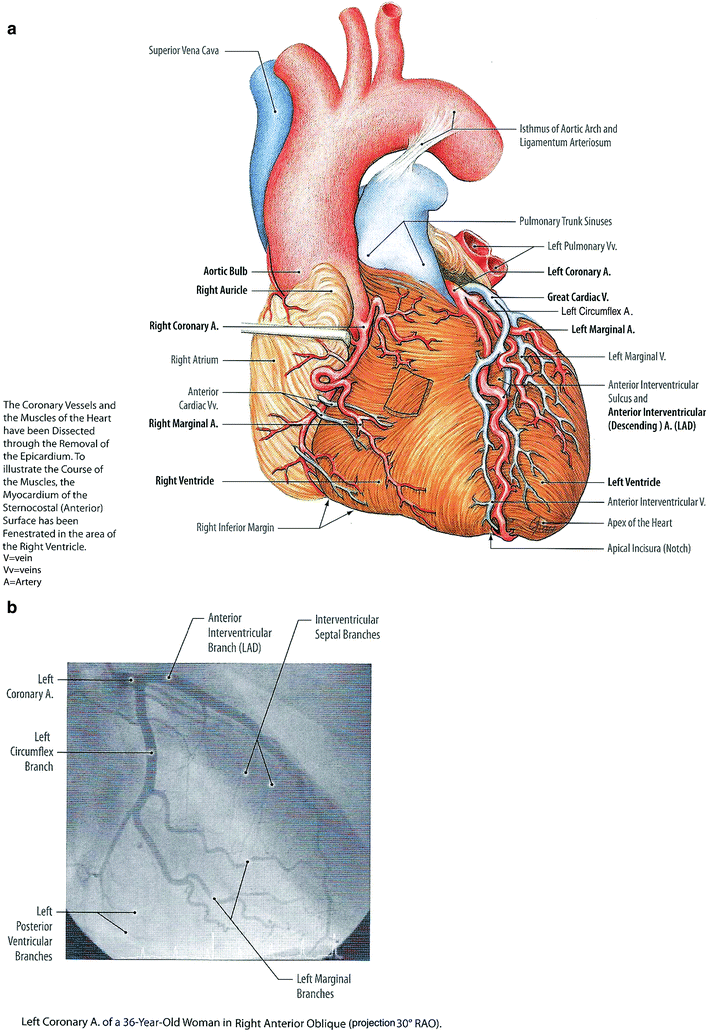
Coronary Arteries Aorta And Great Vessels Arteries And Veins Microcirculation Radiology Key
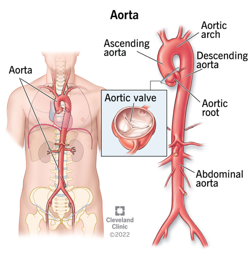
It receives oxygen-rich blood from the heart and distributes it to the body through smaller arteries that branch off of it.
. The superior and petrosal ganglia of the glossopharyngeal nerve are in the jugular foramen. Blood volume is also directly related to blood pressure. Aortic arch The curve of the aorta just after it exits the left ventricle of the heart.
The roots of the ninth and tenth nerves exit together from the medulla and leave the skull through the jugular foramen in the company of the eleventh nerve. C Coarctation of the aorta A problem with the heart. The aorta is the large blood vessel that carries blood from the heart out to the body.
Roll 3 small tubes to represent the arteries that extend from the aorta. The ductus arteriosus is the communication between the pulmonary artery and the proximal descending aorta. Important branches of the abdominal aorta include the arteries that supply blood to the.
Aorta The main artery exiting the heart. Certain lipid-insoluble substances may enter or leave the blood andor pass through the plasma membranes within. The aorta is the largest artery of the body.
In birds the aorta curves to the birds right as it passes dorsal to the heart and toward the backbone but in mammals the aorta curves to the. Attach 3 small 1 4 inch 064 cm tubes to the aorta. The human heart is situated in the mediastinum at the level of thoracic vertebrae T5-T8A double-membraned sac called the pericardium surrounds the heart and attaches to the mediastinum.
A number of age-associated structural changes occur in the arterial system including thickening and dilation of large arteries 44 Figure 3 Table 1. The spleen is found in the left hypochondriac region of the abdomen left upper quadrant. It is used to treat smaller thin infantile hemangiomas.
Its branches distribute oxygenated blood to all parts of the body. Substances can diffuse directly through their plasma membranes if the substances are lipid-soluble. On the first day of life there is a functional closure and an anatomic closure with fibrosis in the first two weeks.
The large artery emerging from the hearts left ventricle that distributes blood to the body. Echocardiographic studies show that the aortic root dilates modestly with age approximating. It shunts blood in utero from the right ventricle to the aorta to bypass the non-functioning lungs.
Blood vessels function to transport bloodIn general arteries and arterioles transport oxygenated blood from the lungs to the body and its organs and veins and venules transport deoxygenated blood from the body to the lungsBlood vessels also circulate blood throughout the circulatory system Oxygen bound to hemoglobin in red blood cells is the most critical nutrient carried by. Arterial elasticity gives rise to the Windkessel effect which through passive contraction after expansion helps to maintain a relatively constant pressure in the arteries despite the. To best depict the location of the spleen well describe its relations.
A valve on the left side of the heart that acts as a one-way gate opening to allow blood to leave the left ventricle and closing to prevent blood from leaking back into that ventricle. A Abnormal arteries in the brain or big blood vessels near the heart. The back surface of the heart lies near the vertebral column and the front surface sits behind the sternum and rib cartilages.
The abdominal aorta is the part of the aorta that passes through the abdominal cavity. The aorta makes up most of the elastic arteries in the body. Medial to the spleen is the left kidney.
Occlusive peripheral arterial disease most commonly develops in the arteries of the legs including the two branches of the aorta iliac arteries and the main arteries of the thighs femoral arteries of the knees popliteal arteries and of the calves tibial and peroneal arteries. It can be applied directly to the hemangioma surface on the skin. If blood pressure within the aorta or the carotid sinus increases the walls of these arteries stretch and stimulate increased activity.
Make sure the small tube runs directly across from the top tube as if both of them make one single piece running through the heart. Superior is the diaphragm while inferiorly it. The nerve descends on the side of the pharynx and then enters the pharynx.
Running through the heart. The upper part of the heart is the attachment point for several large. The aorta springs upward from the left ventricle of heart as the ascending aorta.

Classification Of Aortic Zone Aortic Zone I Extends From The Origin Of Download Scientific Diagram

Surgical Anatomy 1 Ascending Aorta Cannulation Sites On The Ascending Aorta Should Be As High As Safel Anatomy Cardiovascular System Abdominal Aorta

Circulatory System Wikipedia Human Body Activities Human Circulatory System Human Body Systems
Aorta Anatomy And Function
No comments for "Which Arteries Leave Directly From the Aorta"
Post a Comment